Updated Dec 15, 2025
Each year, the School of Medicine and Health Sciences invites SMHS medical students, graduate students, and postdocs to submit their artistic research images to the Art of Science contest. These images showcase our work and passion for science while highlighting the beauty and breadth of research at the SMHS.
Students and postdocs may submit one image that captures their research with artistic style. A jury from SMHS selects winners for cash prizes based on the visual impact of the image.
2026 Winners
We would like to congratulate the 2026 Art of Science Winners! Selected images are on display in the Dean’s Suite in Ross Hall and in Himmelfarb Health Science Library.
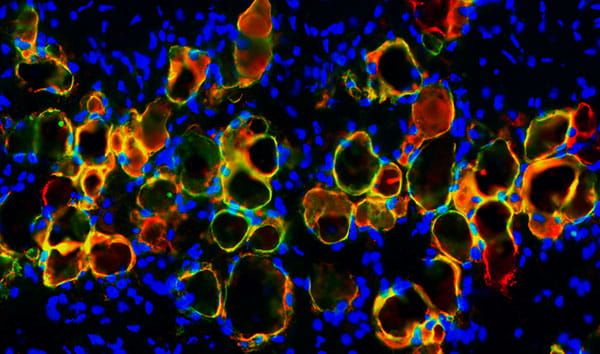
Grand Prize: Sarah Kleb, PhD Student
Advisor: Brett Shook, PhD
Integrated Biomedical Sciences
Adipocyte cells (red) with activated Cre (green)—and co-staining as yellow—in a transgenic model.

Woudasie Admasu, PhD Student
Advisors: Jyoti K. Jaiswal, MSc, PhD and Gustavo Nino, MD
Integrated Biomedical Sciences
Airway epithelial cells express RSV fusion protein (green) at the cell surface (red).

Gabriel Batzli, PhD Student
Advisor: Brett Shook, PhD
Integrated Biomedical Sciences
A Hive plot showing positive-and negative-correlated gene pairs from highly variable genes in skin fibroblasts.

Marcelina Goldstein, Graduate Student
Advisor: Maho Shibata, PhD
Graduate Certificate in Anatomical & Translational Sciences (GCATS) Program
Collagen fibers (red) in human prostate tissue.

Lauren Smith, PhD Student
Advisor: Maho Shibata, PhD
Integrated Biomedical Sciences
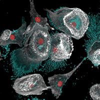
Macrophage immune cells (red) and tumor cells (green) in the prostate duct lumen.
Noah Williams, PhD Student
Advisor: Lauren Woodie, PhD
Integrated Biomedical Sciences
Neurons in the hypothalamus (colors) become active when food is consumed at the wrong time of day.
2025 Winners
|
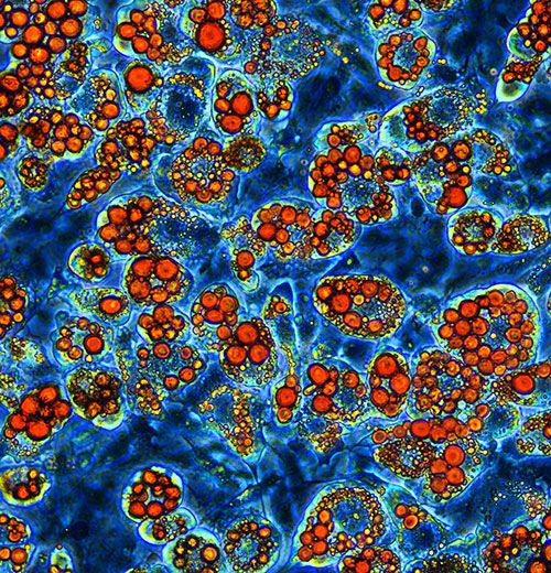
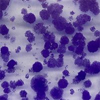
Grand Prize: Sarah Kleb, PhD Student 3T3-L1 fibroblasts that have been differentiated into adipocytes over a 14-day period with serum starvation and a cocktail of insulin, dexamethasone, and IBMX. The newly formed adipocytes have been stained with Oil Red O for analysis of lipid droplet formation within the adipocyte, and rate of differentiation. |
|
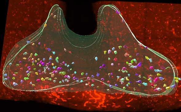
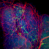
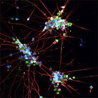
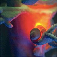
Sally Feng, PhD Student 3-D confocal image of the tip of a prostate lobe from a 5-week-old mouse. Macrophages (Cx3cr1-YFP, green) were found surrounding the prostate in close proximity to nerve fibers (TUBB3, white). |
|
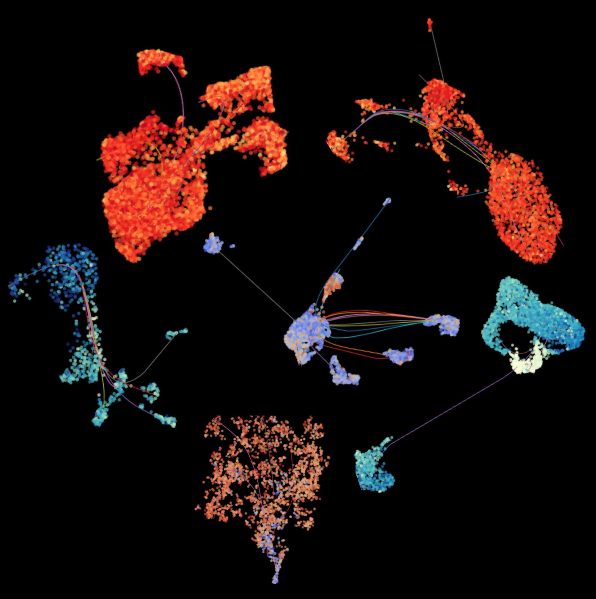
Vania Ballesteros Prieto, MS Student “Sea Creatures.” Dimensionality-reduced visualizations of six single-cell long-read RNA-seq cancer samples generated using scSNViz. |
|
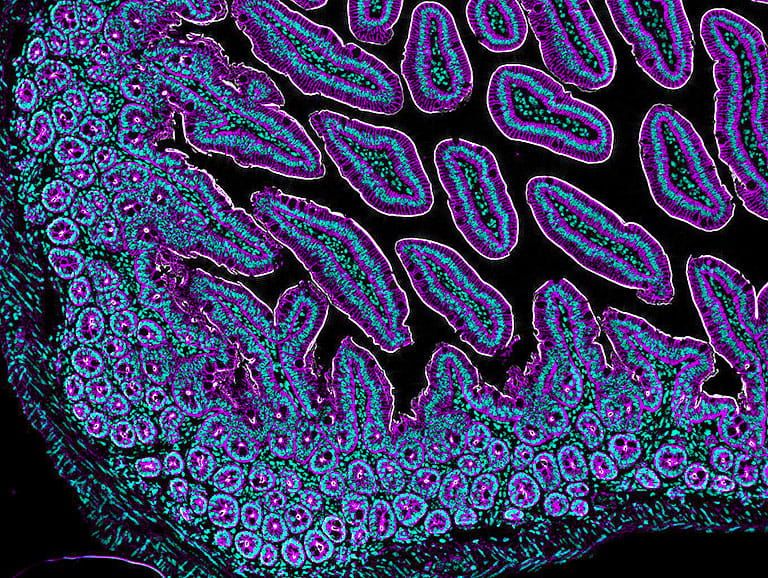
Marta Sanz, Postdoctoral Fellow, and Carles Moreno Soriano, Lab Technician A 20X immunohistochemistry of murine small intestine embedded in paraffin, which highlights cell nuclei stained with DAPI (cyan), the silhouette of enterocytes, goblet cells, and other cells of epithelial origin stained with E-Cadherin (magenta) and the cytoskeletal protein villin which reveals the intestinal brush border (white). The staining is meant to optimize a murine pseudotyped HIV model to study and target mechanisms of viral reservoir establishment. |
2024 Winners
|
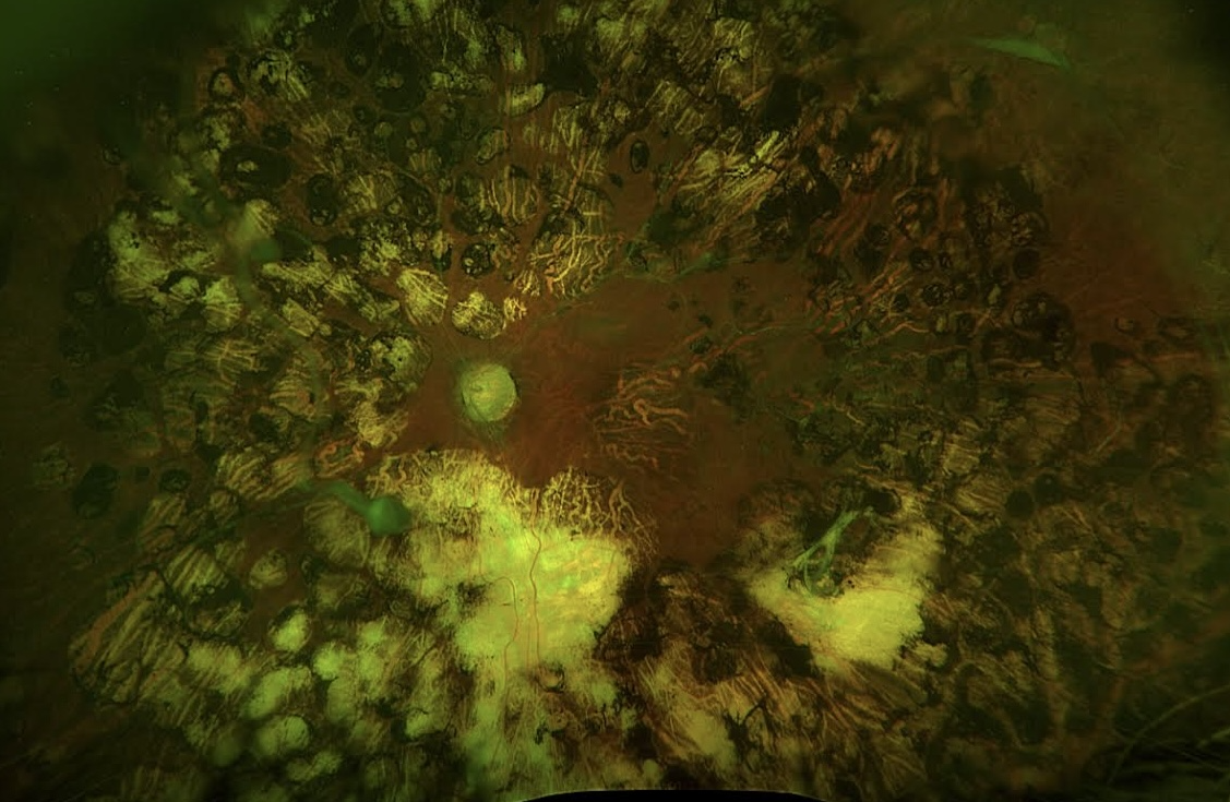
Issa Ali, Medical Student A fundus photo displaying the retina after years of multiple laser scars showing patches or spots that are lighter or darker than the surrounding retinal tissue. These scars are the result of laser photocoagulation therapy, a treatment used to manage retinal conditions such as diabetic retinopathy, retinal breaks, or other retinopathies. |
|
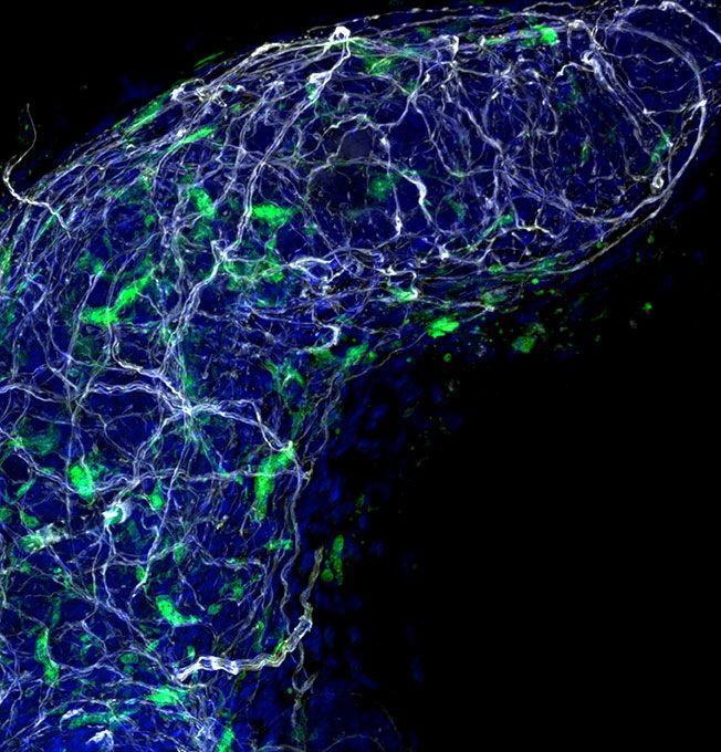
Sally Feng, PhD Student Distal tips of the anterior prostate lobe of a mouse at postnatal day 14 expressing transgenic Csf1r-EGFP (macrophages) in green. Tissue was immunolabeled wholemount for TUBB3 (neurons) in red and stained for DAPI (nuclei) in blue. A maximum intensity projection is shown of the 3D airyscan image. |
|
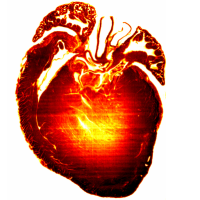
Sarah Tiufekchiev, PhD Student A 2-micron thick section of a D2-mdx dystrophic mouse heart. This slice is one of over 600 sequential images used for our creation of 3D renderings, but represents the midsection, in which you can clearly see areas of fibrosis and large-scale structures of the heart. |
2023 Winners
|
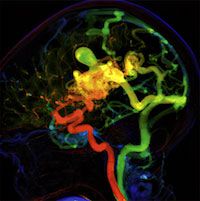
1st Prize: Bennett Levy, Medical Student This is an angiographic parametric image showing a cerebral a arteriovenous malformation with a large intranidal aneurysm (arrow) and a tortuous draining vein (arrow heads). This novel technology is used to understand flow dynamics in real time to help decide on the best treatment strategy. |
|
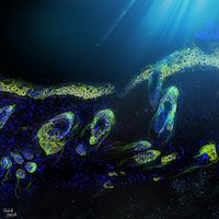
Khatereh Khorsandi, PhD, Postdoc A tiled 20x immunofluorescence image of murine skin, similar to an underwater ocean view. Stained for DAPI (nuclei) in blue, K14 (Keratinocytes) in red, and GFP in green (a mix of both shows a yellow color for keratinocytes), and decorated with the light of the sky plus some lines to display hair follicles that look like jellyfishes. |
|
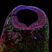
Claire Charpentier, PhD Candidate A mouse cranial neural tube at embryonic day 9.5 imaged using a confocal microscope. All the cells are stained blue (DAPI). The cells from mesodermal origins are green and the neural crest-derived cells are red (transgenic fluorescence). |
|
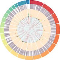
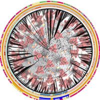
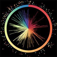
Tyson Dawson, PhD Candidate A radial distribution across chromosomes of key transcripts in Alzheimer's Disease is shown. The overall figure highlights the complex network of expression and regulation of genomic elements that are associated with the progression of Alzheimer's Disease. In the central ring, links are drawn between genomic loci of endogenous retroviruses that have correlated expression and the length and expression levels of these elements are shown in the second and third rings, respectively. |
2021 Winners
|
1st Prize: Hovhannes Arestakesyan, Postdoc 3D reconstruction of astrocyte morphology insubfornical organ of a mouse brain that underwent optical tissue clearing. |
|
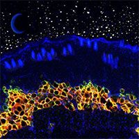
Paula Cooper, Postdoc A tiled 10x immunofluorescence image of murine skin, resembling a landscape. Stained for DAPI (nuclei) in blue, Perilipin1 (adipocytes) in red, and GFP in green, and embellished with a night sky. |
|
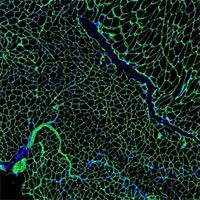
Lana Jagannathan, PhD Student Membrane and nucleus stain of two-year-old wild-type mouse quadricep. |
2020 Winners
|
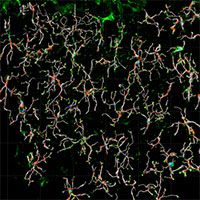
1st Prize: Han Rae Kim, Postdoctoral scientist 3D reconstruction of microglia filaments in the brain subfornical organ. |
|
Tyson Scott Dawson, PhD Student A phylogeny depicting the evolutionary relationships of 2000 SARS-CoV-2. |
|
Tomas Kanholm, PhD Student Fallopian tube cells that have been genetically modified to create a model of ovarian cancer development. |
2019 Winners
|
1st Prize: Naemeh Pourshafie, PhD student Human iPSC-derived motor neurons. Motor neurons are stained with TUJ1(red), HB9(green) and DAPI(blue). |
|
Tyson Scott Dawson, PhD Student Circos plot depicting differentially expressed genes in Systemic Lupus Erythematosus and their location in the human genome. |
|
Cullen Fleming, MD student Images from a negative-space 3D printed model of the ventricular and cisternal system segmented from a brain MRI and labeled with voice-activated LED lights. |
|
Zhe Yu, PhD, Postdoctoral scientist Angiotensin II type 1 receptor-eGFP-BAC mouse was bred with glutamic acid decarboxylase-mCherry mouse and their brain was further stained with anti-parvalbumin antibody. |
2018 Winners |
||
|---|---|---|
|
1st Prize: Brett Eaton, PhD student Marburg virus (turquoise) budding from gray infected dendritic cells (red nuclei) |
|
|
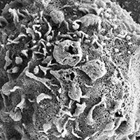
Rachel Burga, PhD student Natural killer cell with iron oxide nanoparticles conjugated to the surface |
|
|
Samantha Dow, PhD student Neurons (red and yellow) derived from apical (blue) and basal (green) progenitor stem cells in the cortex |
|
|
Naemeh Pourshafie, PhD student Motor neuron with dendrites and axons (yellow) and mutant protein aggregates (red) among undifferentiated stem cells. |
|
|
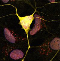
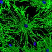
Jessica Schenck, PhD student Astrocytes (green) and nuclei (blue) in the central nervous system |
|
2017 Winners
|
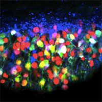
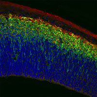
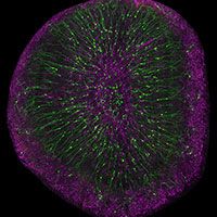
1st Place: Aslam Akhtar, Medical student Olfactory bulb interneurons (green) and astrocytes (magenta) in the brain |
|
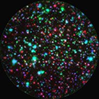
Shabnum Patel, PhD Postdoctoral Fellow T cells producing different cytokines (in different colors) in an immune response to a microbial antigen |
|
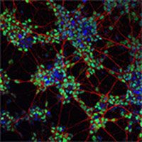
Naemeh Pourshafie, PhD student Spinal motor neurons derived from stem cells with dendrites and axons (red), motor neuron marker (green), and nuclei (blue) |
|
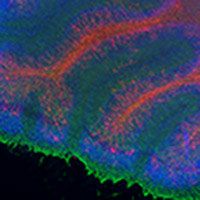
Jessica Schenck, PhD student Cerebellum with myelin (red), astrocytes (green), and nuclei (blue) |
