Art and science may be more closely related than people think, especially when it comes to the beautiful images resulting from scientific research. For a second year, researchers at the George Washington University (GW) School of Medicine and Health Sciences (SMHS) showed off the beauty of the work they do every day.
The Art of Science Contest started last year as a way to show off the dazzling research that medical students, graduate students, and postdoctoral fellows are conducting at SMHS, according to Alison Hall, PhD, associate dean for research workforce development and professor of neurology as SMHS.
This year, more than 17 images were submitted, an increase from last year’s competition, and five entries were chosen as winners.
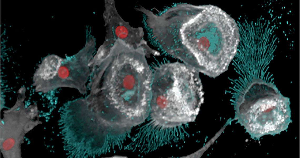
A grand prize of $250 was awarded to Brett Eaton, a PhD student who submitted an image of the Marburg virus budding from infected dendritic cells.
Eaton chose to enter the image, he said, because it had been on his hard drive since 2015 and was an image he always really liked but didn’t think would be used in a publication. “It’s an honor that the panel chose this image,” he said, “but I’m just happy that people will finally get to see it.”
The image, in colors of grey, turquoise, and red, is a microscopic view of the Marburg virus, a close cousin of Ebola, as it attacks the immune system in the early stages of the disease, simultaneously making more of the virus and weakening the body’s ability to fight back, Eaton said.
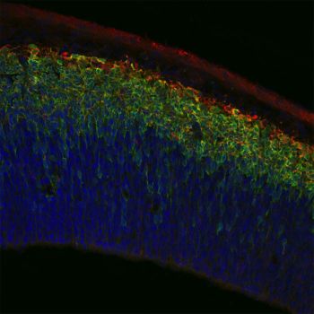
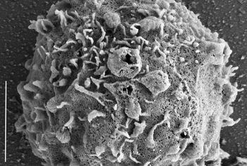
Prizes of $100 were awarded to four other entrants, all GW PhD students. Rachel Burga submitted a scanning electron microscope image of a natural killer cell with iron oxide nanoparticles conjugated to its surface; Samantha Dow’s image featured neurons derived from basal progenitor stem cells in the cortex; Naemeh Pourshafie showed off the beauty of a spinal motor neuron; and Jessica Schenck won with her image of astrocytes in the central nervous system.
The winning images will be displayed in Ross Hall, alongside the winners from last year.


