The George Washington University School of Medicine and Health Sciences (SMHS) and the GW Cancer Center recently received a $600,000 S10 grant from the National Institutes of Health (NIH) to purchase a state-of-the-art BD FACSDiscover™ S8 CellView spectral flow cytometer and live cell sorter. The investment significantly enhances the capabilities of the SMHS Flow Cytometry Core Facility, enabling researchers across disciplines to push the boundaries of biomedical science.
The NIH’s S10 grant program helps fund the purchase of advanced, commercially available research instruments to support the work of NIH-funded investigators. Grants awarded through the program must be shared among multiple researchers, making the program both cost-effective and far-reaching. To qualify, institutions must identify at least three NIH-funded principal investigators with a demonstrated need for the equipment.
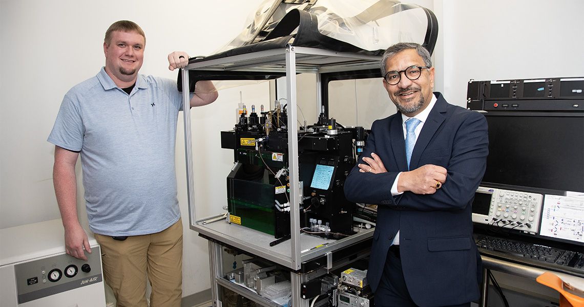
A Leap Forward in Cell Analysis
The new cytometer represents a major step forward for one of the school’s key core lab facilities. Featuring five lasers, 78 fluorescent detectors, and six imaging detectors, the system allows researchers to capture live images of cells and integrate that data into spectral analysis and sorting. This technology offers deeper exploration into cellular traits critical for isolating rare or complex cell populations.
“This instrument is a game-changer,” said Sanjay Maggirwar, PhD, MBA, professor and chair of the Department of Microbiology, Immunology and Tropical Medicine, and PI of the grant application. “It’s the first of its kind to combine high-dimensional spectral analysis with live cell imaging and sorting. We’re not just upgrading equipment; we’re expanding the boundaries of what’s possible in translational research.”
In basic terms, a spectral flow cytometer uses lasers to analyze thousands of cells per second. It detects light from fluorescent markers attached to cells, which emit different colors based on the proteins or characteristics present. This generates a detailed profile of each cell.
According to Gregory Cresswell, PhD, director of the Flow Cytometry Core Facility and coPI on the grant application, the instrument helps researchers understand what specific groups of cells are doing in complex biological environments.
“We work with platelets and immune cells, and follow their interactions. There is a very small population of these cells that interact with platelets,” explained Maggirwar, noting that even a small number of cells can have a large impact on disease. “This complex formation is also very transient, it’s just a kiss and go, but then the immune cells are transformed and they have a pathogenic role in multiple disease conditions.”
By isolating and studying these cells, scientists gain critical insights into how systems in the body function.
Empowering Researchers and Expanding Access
The cytometer will replace the existing equipment and complement the Cytek Aurora analyzer already in use. Together, these instruments will streamline workflows by allowing researchers to directly translate their spectral analysis into live cell sorting.
With its user-friendly software and cloud-based integration, the new system is designed to be accessible to a broad range of users. Even those without technical support from core staff will be able to perform complex experiments independently.
Cresswell emphasized the potential impact, “The new machine will dramatically increase the throughput and flexibility for our users. By removing operational bottlenecks and offering intuitive software, we’re opening the door for more investigators to pursue innovative, high-impact science.”
Alison Hall, PhD, senior associate dean for research, added, “The current cell sorter has been heavily used and is reaching the end of its service contract. It was fantastic that Drs. Maggirwar (Principal Investigator) and Cresswell (Co-Investigator) worked together to submit this grant application. The new equipment provides a state-of-the-art tool for our investigators and will expand our capabilities quite a lot.”
“This modernizes our flow core, and it gives our investigators a competitive edge.”
A Win for Cancer Research and Beyond
The acquisition is fully supported by GW SMHS, with the top-off funds and commitment towards long-term maintenance of the instrument, and will benefit a wide range of NIH-funded investigators. It also marks an important step forward for the GW Cancer Center, especially in fields like immunology, cancer biology, and cellular therapeutics.
Julie Bauman, MD, MPH, Dr. Cyrus Katzen Family Director of the GW Cancer Center, praised the team effort and its broader significance.
“This achievement reflects the extraordinary dedication of Drs. Maggirwar and Cresswell, whose vision and persistence have brought cutting-edge technology to our research community,” she said. “Their work directly supports the GW Cancer Center’s mission to create a cancer-free world through ground-breaking research, innovative education, and equitable care for all. We are proud to celebrate this milestone and the expanded possibilities it brings.”
“In the current environment, getting the funding from NIH for this equipment was a gigantic achievement,” added Maggirwar, who emphasized that the success was a team effort involving both researchers and administrative staff.
“We can’t just rely on the school or the university to provide such expensive resources,” he said. “If we can, we should find alternative funding sources for it.”
Supporting Education and Training
This NIH-funded investment in advanced cytometry technology not only boosts the university's research infrastructure but also expands opportunities for discovery, collaboration, and education. With the new cytometer and cell sorter, GW is positioning itself at the forefront of biomedical innovation — serving both science and society.
Beyond its research applications, the equipment will also enrich educational opportunities at GW. As Maggirwar explained, “We have three NIH T32 training grants funded to GW, as well as several individual F31 grants, that collectively enriches the training outcome for our PhD students; access to such cutting-edge instrumentation adds an extraordinary level of rigor to their research.”


