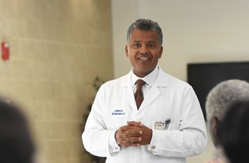
Aiming to accelerate the pace of collaborative clinical and translational research investigations to reduce cancer mortality rates and improve treatment options, George Washington (GW) University’s Cyrus and Myrtle Katzen Cancer Research Center awarded more than $500,000 to 12 university researchers. Now in its fourth year, the Katzen Center, housed within the GW School of Medicine and Health Sciences (SMHS), works in collaboration with GW Hospital and the GW Medical Faculty Associates (MFA). Each year the Katzen Center gives a boost to GW’s cancer research efforts by awarding pilot grants to many of the university’s most promising investigators.
“It’s rewarding to support researchers who are finding clues to why cancers present in patients and identifying cutting-edge therapies for treatment,” said Robert Siegel, M.D., director of the Katzen Cancer Research Center, director of the division of hematology and oncology, and professor of medicine at SMHS.
Robert Hawley, Ph.D., professor and chair of the department of anatomy and regenerative biology at SMHS, along with Imad Tabbara, M.D., professor of medicine at SMHS, will use their $100,000 grant to focus on multiple myeloma. This incurable disease results from the uncontrolled proliferation of the white blood cells that normally produce antibodies. In preclinical studies last year, with the support of the Katzen Cancer Research Center, Hawley and his collaborators developed a test that could be used to determine whether the cancer cells that survive the newest treatment option for multiple myeloma patients exhibit a so-called “multidrug-resistant phenotype.”
Hawley and Tabbara are currently screening multiple myeloma patients to determine whether the assay has prognostic or predictive clinical relevance. “In other words can it be used to monitor how well the therapy is working or help guide treatment decisions,” said Hawley. Success for this team means elucidating the underlying molecular mechanisms that are responsible for the development of drug resistance in multiple myeloma cells that allows them to survive chemotherapy. “A better understanding of drug resistance mechanisms will help guide the future design of more effective therapeutic approaches toward an eventual cure for this devastating disease,” he added.
With their grant, totaling more than $80,000, Samir Agarwal, M.D., assistant professor of surgery at SMHS; and Norman Lee, Ph.D., professor of pharmacology and physiology at SMHS, will sequence the transcriptome (RNA that codes for proteins) and microRNAs (miRNA molecules that regulate gene expression) of cancer specimens. “In this case we are studying colon cancer from African American men and individuals of European descent,” said Lee.
The pair is using next-generation sequencing technology to define differences in the transcriptome of these two racial populations in order to understand why African American men with certain types of cancers have a worst prognosis compared to men of European descent.
“We believe that African American cancers have a set of alternatively spliced mRNAs not found in Caucasian American cancers, and the implication of these alternatively spliced mRNAs that are unique to African American cancers is a more aggressive cancer,” said Lee.
Rachel Brem, M.D., professor of radiology at SMHS, and vice chair of radiology and director of the Breast Imaging and Intervention Center at the MFA, will work with Sidney Fu, M.D., Ph.D., research professor of medicine at SMHS, and use their $100,000 grant for breast cancer research. “The goal of this research is to utilize molecular markers to identify which high-risk lesions found at minimally invasive breast biopsy is associated with cancer,” said Brem. “This will allow fewer women to undergo surgery for diagnosis. We also hope to identify which patients do not need surgery.” According to Brem, this will be the first time the use of molecular markers may result in a decrease in breast surgery for those diagnosed of with breast cancer.
Ajit Kumar, Ph.D., professor of biochemistry and molecular medicine at SMHS and Patricia Latham, M.D., professor of pathology at SMHS, will use their $80,000 grant from the Katzen Cancer Research Center to identify mRNA markers for Hepatitis C virus (HCV) infection associated liver cancer, hepatocellular carcinoma. “We have initiated these studies in a cohort of HCV-infected patients’ sera,” said Kumar.
The team plans to validate the functional impact of miRNA-regulated genes in a mouse model of HCV-infection induced liver tumor to understand how miRNA-targeted genes promote liver tumor in chronic HCV infection. Looking ahead, Kumar and Latham hope to develop molecular markers for early detection of liver cancer and possible novel targets of non-coding RNA based liver cancer therapy.
With their $60,000 grant, Jonathan Sherman, M.D., assistant professor of neurosurgery at SMHS, and director of surgical neuro-oncology, and of stereotactic radiosurgery at the MFA; and Michael Keidar, Ph.D., associate professor of engineering and applied science at the GW School of Engineering and Applied Science (SEAS), proposed a program that introduces cold atmospheric plasma technology in the treatment of malignant brain tumors.
“Plasma is an ionized gas that is typically generated in high-temperature laboratory conditions,” explained Sherman. “Recent progress in atmospheric plasmas has led to a generation of cold atmospheric plasmas (CAP) with ion temperature close to room temperature in the laboratory settings.”
The team’s preliminary data suggests that the CAP pressurized stream jet selectively destroys cancer cells in vitro with less damage to normal cells and significantly reduces tumor size in vivo. Sherman and Keidar envision that this technology could be adapted for clinical use and would fundamentally enhance ablation treatment of solid tumors.
The pair will study CAP in the treatment of glioblastoma, the most aggressive of brain tumors. Their goal is to show that CAP is an effective adjuvant post-surgical treatment option for glioblastoma. “Based on these results we look to develop a new device that can be used intraoperatively to treat malignant brain tumors following surgical resection,” he added.
Kausik Sarkar, Ph.D., associate professor of engineering and applied science at SEAS; and Reza Taheri, M.D., Ph.D., assistant professor of radiology at SMHS, will use their $80,000 to conduct research using microbubbles based ultrasound contrast agents to diagnose thyroid cancer. “Our goal is to make an accurate, cost-effective, and safe diagnostic tool for cancer,” said Sarkar.
The team will study pressure dependent nonlinear ultrasound signals from ultrasound contrast agents. In a malignant nodule, angiogenesis enhances the local hydrostatic pressure. The research aims to use the pressure estimated by the contrast enhanced ultrasound imaging to characterize thyroid nodules and to detect vascular changes associated with malignancy.


