Medical, graduate, and postdoctoral students at the George Washington University (GW) School of Medicine and Health Sciences (SMHS) showcased their passion for biomedical science in the ninth annual Science as Art competition.
Since 2017, students have highlighted the beauty and elegance of ongoing research at GW SMHS, offering a visual window into the complex world of biomedical science. Winners, selected by jury of SMHS faculty, students, and staff, received small cash prizes and will have their images displayed in the Dean’s Suite in Ross Hall.
The 2025 first place winner and three finalists were:
First Prize
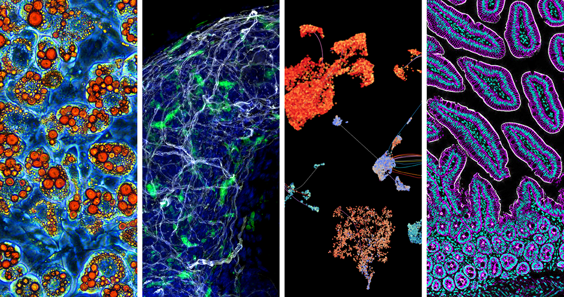
Sarah Kleb, PhD student: 3T3-L1 fibroblasts that have been differentiated into adipocytes over a 14-day period with serum starvation and a cocktail of insulin, dexamethasone, and IBMX. The newly formed adipocytes have been stained with Oil Red O for analysis of lipid droplet formation within the adipocyte, and rate of differentiation.
Finalists
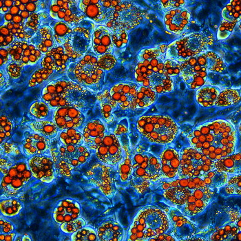
Sally Feng, PhD student: A 3-D confocal image of the tip of a prostate lobe from a 5-week-old mouse. Macrophages (Cx3cr1-YFP, green) were found surrounding the prostate in close proximity to nerve fibers (TUBB3, white).
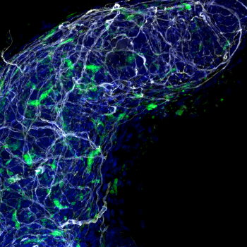
Vania Ballesteros Prieto, MS Bioinformatics & Molecular Biology student: “Sea Creatures.” Dimensionality-reduced visualizations of six single-cell long-read RNA-seq cancer samples generated using scSNViz.
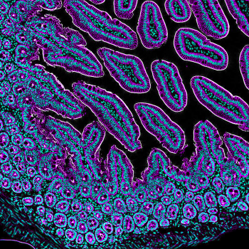
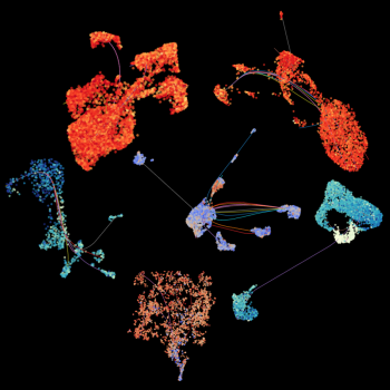
Marta Sanz, postdoctoral fellow, and Carles Moreno Soriano, lab technician: A 20X immunohistochemistry of murine small intestine embedded in paraffin, highlighting cell nuclei stained with DAPI (cyan), the silhouette of enterocytes, goblet cells, and other cells of epithelial origin stained with E-Cadherin (magenta), and the cytoskeletal protein villin, which reveals the intestinal brush border (white). The staining is meant to optimize a murine pseudotyped HIV model to study and target mechanisms of viral reservoir establishment.


